Microtia: Basic Things One Should Know
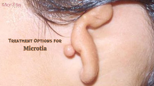
The hearing ability is facilitated by the ears, which also play a significant part in facial attractiveness. External ear deformities and abnormalities are some obvious disorders related to ears and can also impair hearing. Microtia, or little ear, refers to noticeable external ear abnormalities or deformities. It is a congenital disorder in which the ear does not grow properly, resulting in small, oddly shaped, or oftenly absent ears. Both the internal and entire external ear might be impacted by these abnormalities.
Dr. Parag Telang is the founder of "The Microtia Trust," which offers a variety of ear deformity treatments to patients with microtia, including external and internal ear deformities. The expert doctor has shared some crucial insights related to microtia and its related aspects. Every ear surgery is different and it is best to get a professional opinion before moving forward. Keep reading further to find more.
What are the Causes of Microtia?
Although the exact cause of microtia is unknown, environmental and genetic factors are believed to be the primary contributors to this ear condition. The cause of Microtia during fetal development have been explained by several ideas, including disruption of neural crest cells, vascular disturbance, and altitude; however, they have yet to be verified.
Who are the Ideal Candidates for Microtia?
Nine years old and above is the optimal age for performing surgery to repair a nonexistent external ear. Since the regular ear stops growing in size after this age. For Microtia surgery, the rib cartilage must be large enough to build a complete ear framework.
What are the Different Types of Microtia?
- Grade 1: Small ears and an ear canal that is frequently constricted or occasionally absent.
- Grade 2: The upper half of the outer ear is abnormally formed and frequently has a constricted or occasionally absent ear canal.
- Grade 3: The most prevalent ear condition is characterized by small, improperly formed ears without an ear canal.
- Grade 4: The condition with the missing ear and ear canal is called Anotia.
How is Microtia Diagnosed?
Medical professionals typically identify microtia at birth. When the baby is born, the deformity is apparent. A doctor may occasionally do a CT scan as an imaging test to obtain a detailed image of the child's ear. This examination aids them in their search for anomalies in the child's middle and inner ears.
What are the Treatments Available for This?
Although the visual signs of microtia are not always treatable, it is crucial to treat any hearing loss that may be present. Early hearing testing and continuous monitoring of hearing throughout the early infancy stage are crucial. The child can experience speech problems if the hearing loss associated with microtia is not corrected.
Treatment: The cartilage-like soft tissue of the rib is the finest material for creating a new ear. The cartilage for the rebuilt outer ear is taken from the child's rib cartilage. By using the child's tissue, there is significantly less possibility of infection and less probability of rejection. After that, a skin graft is applied over the implant to create a solid, realistic-looking ear.
Note- Artificial materials, like porous polyethylene and silicone, are less desirable options for reconstructed ears since they are susceptible to early and late rejections, infection, and exposure.
How Many Stages are there in Microtia Surgery?
Two stages are involved in microtia surgery:
- The rib tissue or cartilage is taken in the initial stage. Harvested cartilage is used to build a three-dimensional scaffolding for the ear. The prosthetic ear is made from cartilage using the patient's other healthy ear as a template. The entire ear framework is then built and placed in the proper location on the side of the head.
- From the side, the ear appears complete, but the lobe is absent. Between three and six months following the first stage, the second stage is when the lobe behind the ear is formed. For 5-7 days, a tight bandage keeps the skin graft in place.
What are the Risks Involved?
- Scarring: They are permanent but are hidden behind the ear
- Scar Contraction: As they heal, surgical scars may tighten and harm the skin surrounding the ear.
- Skin Breakdown: The skin that covers the ear structure may deteriorate, revealing the implant or cartilage below.
- Damage to the Graft Site: Scars may appear where the skin was removed from another body region to construct a flap to cover the ear framework.
What are the Benefits of this?
- Using the patient's rib cartilage eliminates the possibility of graft rejection.
- Natural-looking results
- Improves the balance and look of the face.
- It aids in people gaining self-assurance and respect.
All these factors mentioned above will be helpful for one to decide whether to go for ear surgery or not. But one can visit The Microtia Trust for learning about the Ear Surgery Cost in Mumbai and get a consultation from a skilled doctor, Dr. Parag Telang to get a clearer picture and a thorough look at all the ways to treat ear deformities.


Comments
Post a Comment