What is Microtia? Causes, Types and Treatment
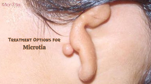
The ears help with hearing and contribute significantly to one’s facial appearance. Some visible ear problems affecting hearing include outer ear deformities and abnormalities. Microtia, or "little ear," describes observable abnormalities or deformities of the external ear. It is mostly termed as a genetic condition where the ears do not develop normally, leaving them small, abnormally shaped, or frequently absent. These anomalies may affect both the inner and entire outer ear.
At “The Microtia Trust," founded by Dr. Parag Telang, the Best Plastic Surgeon in Mumbai, provides patients with various ear deformity therapies for outer and inner ear abnormalities. To learn more, continue reading.
What is Microtia?
A congenital condition called microtia causes the auricle (outer ear) to be undeveloped. The term "anotia" refers to a pinna that has not yet fully grown. It is described as microtia-anotia since microtia and anotia share the exact origin. Bilateral or unilateral microtia are both possible (affecting both sides).
What are the Causes of Microtia?
The reason for microtia in children is yet unknown, but some examples link the condition to genetic flaws in several or just one gene, elevation, and gestational diabetes. Babies delivered underweight, male sex, women's gravidity, parity, and medicines used during pregnancy are risk factors identified by research. Although it has not been well investigated, genetic inheritance has been found to happen in the first few months of pregnancy in the few research that has been done.
Which Candidates can undergo Microtia Surgery?
Children with completely formed ears and rib cartilage that is sufficiently thick and strong to develop an adult-sized ear are typically between the ages of 9 and 15 years old.
Individuals in good health with reasonable aspirations.
What are the Types of Microtia?
Grade 1: Little ears with a typically narrowed or occasionally missing ear canal.
Grade 2: The ear canal is usually constricted or missing in the top portion of the outer ear, which is improperly constructed.
Grade 3: Small, poorly shaped ears lacking an ear canal are the most common type of ear abnormality.
Grade 4: Anotia is when one ear and the ear canal are absent.
How is Microtia Treated?
Under general anesthesia, rib cartilage is used as a graft in microtia ear surgery, so the child won't experience any pain. The steps involved are as follows:
Harvesting of rib cartilage graft: A 1-1.5 inch long incision is made over the child's sixth, seventh, and eighth ribs in the chest, and a portion of the floating ribs with rib cartilage is removed.
Creation of the cartilage framework for the new ear: The structure for the new ear is created by painstakingly carving and piecing together the removed rib cartilage. Each component of the replacement ear framework is built to order and put together to match the patient's other ear. Copies of the other normal ear that were manufactured using 3D technology were used to assist in building the ear framework.
Placing of the cartilage ear framework at the side of the head: When the ear structure is finished, it is placed inside a skin pocket that has been made just below the scalp at the side of the face, wherein the ear must be recognized. The inferior fleshy portion of the ear, known as the lobule, is created using the patient's own ear remains. The ear cartilage structure then integrates into the patient's live tissues during the next three to four months throughout the healing process.
Fine-tuning of reconstructed ear and smoothing out of earlobe: Additional surgeries could be necessary to finish ear reconstruction after the ear has healed. This entails making a few modifications to the rebuilt ear. This is microtia surgery's second stage. To remove the ear from the head tissue at this stage, the surgeon creates incisions behind the ear. The cartilage structure is then raised, allowing the ear to "pop out" or expand and match the other side. The freshly elevated ear is covered on the rear so that it can be lifted away from the skull using skin and cartilage grafts. Additional minor procedures may be performed in some instances to enhance ear shape, minimize the visibility of scars, or increase ear protrusion.
What Happens After the Surgery?
After the procedure, the patient can experience some chest and ear discomfort. Incision sites may have surgical drains implanted to relieve pressure and edema and lessen the collection of blood or even other fluids. The patient's new ear is covered with a light dressing or a postoperative ear protection to safeguard the repaired ear for approximately 10 to 14 days.
One can visit The Microtia Trust to learn about the Ear Plastic Surgery Cost in Mumbai and get a consultation from a skilled doctor, Dr. Parag Telang, to get a clearer picture and a thorough look at all the ways to treat ear deformities.


Comments
Post a Comment